Feline Cutaneous Hemangioma: Blood Vessel Skin Tumor
1. Why this topic matters to cat owners
Finding a new bump, red spot, or “blood blister” on your cat’s skin can be unsettling—especially when it seems to appear quickly or bleeds easily. While many skin growths in cats are benign (non-cancerous), some need prompt attention to prevent discomfort, infection, or progression to more serious disease.
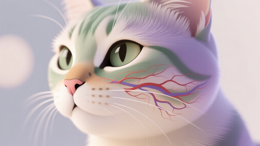
One condition that can look dramatic but is often treatable is a feline cutaneous hemangioma. This is a tumor made up of blood vessels in the skin. It may look like a small red-to-purple mass or a bruise-like area. Because it can resemble other skin tumors (including malignant ones), the safest approach is always to have any new or changing skin lesion evaluated by a veterinarian.
2. Overview: what is a cutaneous hemangioma?
A cutaneous hemangioma is a benign tumor formed from blood vessel cells. “Cutaneous” means it’s in the skin. “Hema” refers to blood, and “angioma” refers to a tumor arising from blood vessels.
These growths typically:
- Develop in the skin (and sometimes just under it)
- Contain clusters of small blood vessels
- May look red, purple, or dark due to blood within the lesion
- Can bleed if scratched, rubbed, or traumatized
Hemangiomas are generally considered non-cancerous. However, they can be confused with other conditions that may look similar, including:
- Hemangiosarcoma (a malignant blood-vessel tumor)
- Mast cell tumors
- Abscesses or infected cysts
- Hematomas (collections of blood under the skin)
- Other benign skin masses (like cysts or papillomas)
That’s why diagnosis matters: the appearance alone can’t reliably tell you which type of lesion it is.
3. Symptoms and warning signs to watch for
Some cats act completely normal and the only sign is the growth itself. Others may show irritation if the area is tender or frequently bleeds.
Common signs cat owners notice
- A small red, purple, or bluish bump on the skin
- Bruise-like discoloration that doesn’t resolve as expected
- A raised “button” mass that may be smooth or slightly irregular
- Bleeding or oozing, especially after scratching or grooming
- Scabbing that keeps recurring in the same spot
- Hair loss over or around the lesion
Changes that should prompt a quicker vet visit
- Rapid growth over days to weeks
- Repeated bleeding (spots of blood on bedding, collars, or your hands)
- Ulceration (an open sore) or a foul odor
- Multiple new lesions appearing
- Swelling or redness around the mass (possible infection or inflammation)
Practical at-home actions you can do today
- Measure it (in millimeters) and write it down.
- Take a clear photo next to a coin or ruler once a week.
- Prevent self-trauma if it’s being scratched: ask your vet about an e-collar or soft recovery collar.
- Avoid human ointments unless your vet recommends them—some are unsafe if licked.
4. Causes and risk factors
In many cats, the exact cause of a hemangioma isn’t known. That said, veterinarians have identified factors that may increase risk.
Possible risk factors
- Sun exposure (UV light): Cats with light-colored fur or thin hair coats may be more prone to sun-related skin damage, particularly on sparsely haired areas.
- Light pigmentation: White or pale-coated cats can be more vulnerable to UV-associated skin problems.
- Age: Skin tumors are more common in middle-aged to older cats, though benign lesions can occur at various ages.
- Chronic irritation or trauma: Repeated rubbing (collars, harnesses) or scratching may aggravate lesions and make them more noticeable.
Hemangiomas can occur on different body areas, but lesions may be more noticeable on areas with thinner fur or more sun exposure (such as the head, ears, or trunk). Any location is possible.
5. Diagnosis: what to expect at the vet
A veterinary visit is the best next step for any new skin mass. The main goal is to identify the type of growth and rule out malignant or infectious conditions.
Typical diagnostic steps
- Full physical exam with careful skin assessment (including checking for additional lumps).
- History questions: How long it’s been there, any bleeding, changes in size, indoor/outdoor status, sunbathing habits, medications, and overall health.
- Fine needle aspirate (FNA): A small needle collects cells for microscopic evaluation. With blood-vessel tumors, the sample can sometimes be very bloody and may not provide a clear answer.
- Biopsy: If the mass is suspicious, changing, bleeding, or the FNA is inconclusive, the vet may recommend removing part (incisional biopsy) or all (excisional biopsy) and sending it to a pathologist.
- Histopathology: This lab analysis is the gold standard to confirm hemangioma and distinguish it from hemangiosarcoma.
Additional tests your vet may recommend
- Bloodwork (CBC/chemistry) before anesthesia or surgery, especially in older cats.
- Imaging (less common for a simple skin hemangioma) if there is concern about deeper involvement or another condition.
How to prepare for the appointment
- Bring your photos and measurements.
- List any medications, supplements, or flea/tick products.
- Prevent grooming of the lesion beforehand if it tends to bleed.
6. Treatment options (medical, surgical, home care)
Treatment depends on size, location, bleeding, and what the pathology report shows. Because hemangiomas can mimic malignant tumors, your veterinarian will often recommend removal or biopsy rather than “watching” indefinitely.
Surgical options
- Surgical excision (removal): Often the treatment of choice. Many hemangiomas are cured with complete removal.
- Margins: Your vet will aim to remove the mass with a small rim of normal tissue when appropriate. The pathologist reports whether the margins are clean.
- Laser surgery (where available): May reduce bleeding and be helpful for certain lesions or locations.
Medical management
There is no single “pill” that reliably eliminates a cutaneous hemangioma in cats. Medical treatment is generally supportive, used when surgery is not immediately possible or when managing complications.
- Pain relief: Your vet may prescribe cat-safe pain medication after biopsy or removal. Never give human pain relievers.
- Antibiotics: Only if there is evidence of infection (redness, discharge, odor) or after certain procedures.
- Topical care: Sometimes recommended to protect the surface or support healing, but only use products approved by your veterinarian.
Home care after surgery or biopsy
- E-collar or recovery collar: Prevents licking and chewing, which is one of the most common causes of incision problems.
- Keep the incision clean and dry: Avoid bathing until your vet clears it.
- Monitor daily: Look for swelling, heat, discharge, missing sutures, or worsening redness.
- Activity restriction: Limit jumping and rough play during healing.
- Give medications exactly as directed: If your cat seems painful or overly sleepy, call your vet for guidance.
7. Prevention strategies and early detection tips
You can’t prevent every skin tumor, but you can reduce risk and catch problems early.
Ways to reduce risk (especially sun-related)
- Limit sunbathing during peak hours (late morning through afternoon), particularly for white or lightly pigmented cats.
- Create shaded resting spots near windows or use UV-filtering window film/screens.
- Keep cats indoors or provide supervised outdoor time to reduce UV exposure and skin injuries.
Early detection habits that help
- Monthly “nose-to-tail” checks: Feel for bumps when petting and brushing.
- Check common hidden zones: Under the chin, armpits, groin, belly, between toes, and around the tail base.
- Track any lump for 30 days: If it persists, grows, bleeds, or changes color, schedule a veterinary exam sooner rather than later.
Any lump that is new, changing, or bleeding deserves professional evaluation—even if your cat seems otherwise fine.
8. Prognosis and quality of life considerations
The outlook for a cat with a confirmed cutaneous hemangioma is often very good, especially when the lesion is fully removed. Many cats return to normal quickly after minor surgery, and the growth does not come back at the same site when excision is complete.
Quality of life considerations include:
- Comfort: Lesions that bleed or scab can be irritating; removal often improves comfort.
- Location challenges: Tumors on areas like the legs, face, or thin-skinned regions may require extra planning for closure and aftercare.
- Monitoring: Even after a benign diagnosis, keep doing regular skin checks. Cats can develop unrelated skin growths over time.
If pathology reveals a different diagnosis (such as a malignant tumor), your veterinarian will discuss staging tests and a tailored treatment plan. Getting an accurate diagnosis early gives you the best options.
9. When to seek emergency veterinary care
A hemangioma isn’t usually an emergency by itself, but complications can be urgent. Seek prompt or emergency care if you notice:
- Bleeding that won’t stop within 10–15 minutes of gentle pressure with clean gauze or a soft cloth
- Sudden swelling of the mass (possible bleeding into the lesion)
- Pale gums, weakness, collapse, or rapid breathing (signs of significant blood loss or another serious problem)
- A rapidly enlarging mass accompanied by pain
- Signs of infection: pus-like discharge, foul odor, fever, or the area is hot and very painful
- Post-surgical concerns: incision opens, heavy bleeding, or your cat is lethargic and won’t eat after surgery
If you’re unsure, call your veterinary clinic or an emergency hospital—they can help you decide the safest next step.
10. FAQ: common questions cat owners ask
Is a cutaneous hemangioma in cats cancerous?
A hemangioma is typically benign (not cancer). The challenge is that other tumors—like hemangiosarcoma—can look similar, so a veterinarian often recommends biopsy or removal to confirm the diagnosis.
Can a hemangioma bleed a lot?
It can bleed more easily than many other skin bumps because it’s made of blood vessels. Minor bleeding is common if the surface is scratched, but persistent or heavy bleeding should be treated as urgent and evaluated by a veterinarian.
Should I wait and watch the lump for a while?
It’s reasonable to monitor a very small, stable lump for a short period if your veterinarian agrees. For most cats, though, the safest plan is to schedule an exam soon, especially if the lesion is new, changing, or bleeding. Photos and measurements help your vet assess progression.
How is hemangioma different from a “blood blister” or hematoma?
A hematoma is a collection of blood under the skin, often from trauma, and it may resolve over time. A hemangioma is a true growth of blood-vessel tissue and typically won’t disappear on its own. A vet exam and diagnostic testing are needed to tell them apart.
Will my cat need surgery?
Many cats do best with surgical removal, especially if the lesion bleeds, grows, or the diagnosis is uncertain. Surgery is often curative for hemangiomas, and your veterinarian can discuss anesthesia safety and aftercare based on your cat’s age and health.
Can I put antibiotic ointment or a bandage on it at home?
It’s best not to apply human products without veterinary guidance—cats lick, and some ingredients are harmful if swallowed. Bandages can also cause rubbing or moisture buildup. If the lesion is bleeding, gentle pressure with clean gauze is appropriate, then contact your veterinarian for next steps.
If you’ve found a suspicious skin spot or lump on your cat, a veterinary check is the most reassuring and effective way to get answers. For more cat health articles, practical wellness tips, and guidance you can use at home, visit catloversbase.com.