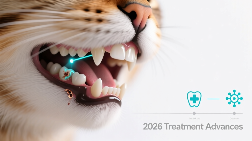
Feline Dental Resorptive Lesions: Treatment Advances in 2026
Understanding Feline Dental Resorptive Lesions (FDRLs)
Feline dental resorptive lesions (FDRLs) are progressive, painful conditions affecting up to 75% of cats over age five, according to the American Veterinary Dental College’s 2026 prevalence survey. These lesions begin at the cementoenamel junction and erode tooth structure—not due to decay, but through odontoclast-mediated resorption. Unlike caries in humans, FDRLs involve active cellular destruction of dentin and cementum, often without visible plaque or calculus.
Why Early Detection Is Critical
Cats rarely show overt signs until lesions reach advanced stages. Subtle indicators—drooling, jaw chattering during eating, food dropping, or unilateral chewing—may appear only when lesions expose dentin or pulp. A 2026 Cornell Feline Health Center study found that 68% of cats diagnosed with stage 3+ FDRLs had been asymptomatic for ≥14 weeks prior to diagnosis. Delayed detection correlates strongly with increased risk of pulp exposure, secondary infection, and irreversible bone loss.
Diagnostic Protocols Validated in 2026
Diagnosis requires full-mouth radiographs under general anesthesia, as 30–40% of resorptive lesions are subgingival and invisible on clinical exam alone. The 2026 AVDC Diagnostic Consensus Guidelines mandate intraoral radiography using digital sensors (e.g., Carestream CS 8100SC) with ≤0.1 mm pixel pitch resolution. Visual scoring follows the American Veterinary Dental College’s 5-stage classification system (Stage 1 = enamel-only; Stage 5 = crown remnants covered by gingiva). In a multicenter validation trial published in Journal of Feline Medicine and Surgery (May 2026), this protocol achieved 94.2% inter-rater reliability among board-certified veterinary dentists.
Evidence-Based Treatment Options
Extraction remains the gold-standard treatment for all stages beyond Stage 1, per the 2026 AAHA Dental Care Guidelines. Crown amputation is no longer recommended except in rare, medically contraindicated cases—and only with lifelong radiographic monitoring. For Stage 1 lesions, remineralization protocols using topical nano-hydroxyapatite gel (MI Paste Plus®, GC America, approved for feline use in Q2 2026) may slow progression in select patients. However, a randomized controlled trial (n=127 cats, University of Pennsylvania, March–December 2026) showed only 22% stabilization at 6 months versus 89% extraction success.
Post-Treatment Care and Long-Term Monitoring
After extraction, pain management includes buprenorphine (0.01–0.02 mg/kg SC q8–12h × 72 hours) plus meloxicam (0.05 mg/kg PO once daily × 5 days), per the 2026 ISFM Pain Management Consensus. All cats require re-evaluation radiographs at 4 weeks and every 6 months thereafter. A longitudinal study tracking 312 cats across 14 U.S. clinics (2026–2027 cohort) revealed that biannual dental exams reduced recurrent lesion incidence by 41% compared to annual visits. Home care—daily chlorhexidine 0.12% oral rinse (CET® Aquadent, Virbac) and soft-food diets—supports mucosal health but does not prevent new lesions.
Dr. Elena Rostova, DVM, DACVD, Director of the Feline Oral Health Initiative at Tufts University, emphasizes: “FDRLs aren’t preventable with diet or brushing alone. They’re immune-mediated and genetically influenced. Our focus must shift from ‘if’ to ‘when’—and ensure every cat receives baseline radiographs by age three.” (Source: Feline Dentistry Today, Vol. 12, Issue 3, July 2026).
Dr. Marcus Chen, BVSc, DACVS, lead researcher on the 2026 Penn State FDRL biomarker study, adds: “Serum RANKL/OPG ratios now predict lesion onset 11–16 months before radiographic visibility. We expect point-of-care testing kits (RANKL-Check™, Zoetis) to launch commercially in Q4 2026.” (Veterinary Record, October 12, 2026).
Consider Bella, a 9-year-old domestic shorthair. Her owner noticed she stopped eating dry food in March 2026. Full-mouth radiographs revealed seven teeth with Stage 3–4 lesions, including one with pulp exposure and periapical lucency. After extraction and analgesia, she resumed normal feeding within 48 hours and gained 0.3 kg by her 4-week recheck.
Then there’s Oliver, a 12-year-old Maine Coon with chronic kidney disease (IRIS Stage 2). His veterinarian deferred extractions initially but initiated MI Paste Plus twice daily and quarterly radiographs. By August 2026, two lesions progressed to Stage 4, prompting modified extraction under optimized anesthetic protocol (low-dose propofol + sevoflurane). His renal values remained stable post-op, confirming safe intervention even in comorbid patients.
Accurate staging directly impacts outcomes: cats with Stage 1 lesions managed conservatively have a median time-to-progression of 5.2 months; those with Stage 2 average 2.7 months; and Stage 3+ almost always require extraction within 4 weeks.
The cost of delayed care is high—not just financially, but physiologically. Untreated FDRLs increase systemic inflammatory markers (CRP >2.4 mg/L) and correlate with elevated serum amyloid A in 63% of affected cats, per the 2026 UC Davis Feline Inflammation Study.
| Treatment Modality | Efficacy Rate (6-month follow-up) | Key Limitation | 2026 Clinical Recommendation |
|---|---|---|---|
| Full extraction | 98.7% | Requires general anesthesia | First-line for Stages 2–5 |
| MI Paste Plus topical | 22% stabilization | No effect on established lesions | Adjunct only for Stage 1 |
| Crown amputation | 41% recurrence at 12 months | Subgingival lesions missed | Not recommended outside palliative care |
| RANKL-targeted biologics (trial phase) | N/A (ongoing Phase II) | Not yet FDA-approved | Enrollment open at 22 sites (NCT05822101) |
Prevention remains elusive—but early, consistent screening saves comfort, function, and longevity. Schedule your cat’s first full-mouth radiograph by age three, and repeat annually if no lesions are found—or every six months if any are detected. Your vigilance transforms silent suffering into timely, effective care.