Cat Corneal Dystrophy: Inherited Eye Surface Clouding
1. Introduction: Why This Topic Matters to Cat Owners
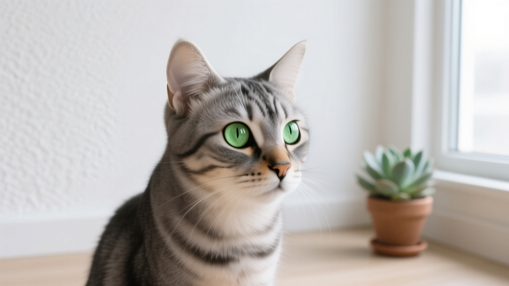
Your cat’s eyes do a lot of quiet work every day—tracking movement, navigating dim rooms, and communicating mood. When a normally clear eye starts to look cloudy or develops a white spot, it’s understandable to worry about pain, vision loss, or an infection. One possible cause is corneal dystrophy, a group of inherited conditions that affect the clear “window” at the front of the eye.
Corneal dystrophy is often non-infectious and not caused by anything you did. Many cats feel fine and keep living normal lives, but any change to the eye deserves a veterinary evaluation. Some eye problems look similar on the surface yet require very different treatment. Knowing the basics helps you catch changes early, get the right care quickly, and protect your cat’s comfort and vision.
2. Overview: What Is Corneal Dystrophy in Cats?
The cornea is the clear, curved surface covering the front of the eye. It’s made of layers, a bit like a multi-layered windshield:
- Epithelium (outer “skin” layer)
- Stroma (the thick, supportive middle layer)
- Descemet’s membrane and endothelium (inner layers that help keep the cornea clear by managing fluid)
Corneal dystrophy describes inherited abnormalities where one of these layers develops changes that lead to cloudiness or opacity. Depending on which layer is affected, the cloudiness may look like:
- A white, gray, or bluish haze
- Crystalline or “sparkly” deposits
- Well-defined circular spots or more diffuse clouding
Corneal dystrophy is typically:
- Bilateral (affects both eyes), though one eye may look worse at first
- Slowly progressive or stable over time
- Non-inflammatory early on (meaning the eye may not be red or painful)
That said, corneal dystrophy can sometimes lead to complications like irritation or ulceration, which can be painful. This is one reason eye clouding should never be “watch and wait” without professional guidance.
3. Symptoms and Warning Signs to Watch For
Corneal dystrophy itself may be subtle at first. You might notice changes during a sunny window nap or when your cat turns toward a light.
Common signs owners notice:
- White, gray, or bluish cloudiness on the eye surface
- One or more opaque spots on the cornea
- Both eyes affected (sometimes unevenly)
- Eyes that otherwise look “quiet” (little to no redness early on)
Signs that suggest discomfort or a complication (needs prompt vet attention):
- Squinting or holding an eye closed
- Increased blinking
- Pawing at the face or rubbing the eye on furniture/carpet
- Tearing or watery discharge
- Thick yellow/green discharge
- Redness of the white part of the eye
- Light sensitivity (hiding from bright areas)
- Behavior changes: irritability, reduced appetite, less play
At-home check you can do today: In a well-lit room, gently look at both eyes from the front and side. Note whether the cloudiness is in the same location on both eyes, whether it seems on the surface vs deeper, and whether there’s redness or discharge. Take a clear photo (no flash if possible). Photos help your veterinarian track subtle changes over time.
4. Causes and Risk Factors
Corneal dystrophy is considered inherited (genetic), meaning it’s related to how the cornea’s cells develop and maintain clarity. It is not contagious to other pets or people.
Potential risk factors and patterns:
- Genetics/breed predisposition: Corneal dystrophies are well recognized in some purebred populations; cats can be affected too, though feline cases are less commonly discussed than canine ones.
- Age of onset: Often noticed in young to middle-aged cats, though it can be detected at any age depending on severity and visibility.
- Family history: Related cats may show similar corneal changes.
Conditions that can look similar but are not corneal dystrophy:
- Corneal ulcer (scratch/injury)
- Corneal sequestrum (more common in cats; dark brown/black corneal plaque)
- Chronic inflammation (keratitis)
- Corneal scarring from past trauma or infection
- Glaucoma (may cause a bluish, “steamy” appearance with pain)
- Cataract (cloudiness inside the eye lens, not on the surface)
- Systemic issues causing lipid deposits (less common, but considered)
This overlap is why a veterinary eye exam is so valuable—two conditions can look similar to you at home but require very different care.
5. Diagnosis: What to Expect at the Vet
Your veterinarian will focus on two big questions: What layer is affected? and Is the eye painful or at risk?
A typical diagnostic workup may include:
- History and timeline: When you first noticed clouding, whether it changed quickly, and any squinting or discharge.
- Full eye exam: Using bright light and magnification to examine the cornea’s surface and depth.
- Fluorescein stain: A safe dye that highlights corneal ulcers or scratches (ulcers often glow green under blue light).
- Tear production testing (Schirmer tear test): Less common in cats than dogs, but may be used if dry eye is suspected.
- Intraocular pressure measurement (tonometry): Helps screen for glaucoma or uveitis.
- Ocular ultrasound (if the vet can’t see deeper structures well) to assess inside the eye.
- Referral to a veterinary ophthalmologist: Recommended if the diagnosis is uncertain, vision is threatened, or advanced treatments are being considered.
What you can bring to the appointment:
- Photos of your cat’s eyes over time
- A list of any medications (including human eye drops—do not apply them unless directed)
- Notes about behavior changes, appetite, squinting, or rubbing
6. Treatment Options: Medical, Surgical, and Home Care
Treatment depends on whether the corneal dystrophy is causing discomfort, affecting vision, or triggering secondary problems. Some cats require little to no treatment beyond monitoring, while others need supportive care.
Medical Management
- Lubricating eye drops/gel: Helps protect the corneal surface and improve comfort if mild irritation is present. Use only products recommended by your veterinarian—cats can react to preservatives in some formulations.
- Topical antibiotics: Not for dystrophy itself, but commonly prescribed if there’s a corneal ulcer or high risk of surface infection.
- Pain control: If the eye is painful, your vet may prescribe systemic pain relief. Avoid giving human pain medications—many are dangerous for cats.
- Anti-inflammatory medications: Only when appropriate and under veterinary supervision. Some anti-inflammatory drops are unsafe if an ulcer is present, which is why staining is crucial.
Surgical/Advanced Options
Surgery is not needed for most cases, but may be discussed when there are repeated ulcers, significant surface irregularity, or vision-threatening changes.
- Superficial keratectomy: A procedure where abnormal superficial corneal tissue is removed. Often performed by a veterinary ophthalmologist.
- Corneal grafting or advanced repair: Considered if there is deep ulceration or severe corneal damage (more related to complications than dystrophy alone).
Home Care You Can Do Safely
- Prevent rubbing: If your cat is pawing at the eye, use an e-collar (cone) as directed and contact your vet.
- Reduce irritants: Avoid dusty litter, strong fragrances, aerosol cleaners, smoke, and windy car rides with open windows.
- Give medications exactly as prescribed: Eye medication schedules matter. Set phone reminders.
- Monitor daily during flare-ups: Watch for squinting, discharge, or worsening cloudiness.
Do not do these at home:
- Do not use leftover eye meds from another pet.
- Do not use human “redness relief” drops.
- Do not try to wipe the cornea directly; if discharge is present, clean only the fur around the eye with a damp cotton pad.
7. Prevention Strategies and Early Detection Tips
You can’t prevent the genetic tendency for corneal dystrophy, but you can prevent avoidable complications and catch changes early.
- Schedule routine wellness exams: Ask your veterinarian to include an eye check at every visit, especially if you’ve noticed any clouding.
- Track changes with photos: A monthly photo in the same lighting can reveal slow progression.
- Protect the eye surface: Promptly address squinting, redness, or discharge—these signs often indicate irritation or ulceration, which needs treatment.
- Keep nails trimmed: Overly sharp nails increase the risk of self-inflicted corneal scratches.
- Safe play: Avoid toys with sharp edges and supervise play that could lead to eye trauma.
- Breeding considerations: If your cat is a breeding animal and diagnosed with an inherited eye condition, discuss ethical breeding choices with your veterinarian or a veterinary ophthalmologist.
8. Prognosis and Quality of Life
The outlook for cats with corneal dystrophy is often good, especially when the condition is mild and non-painful. Some cats have stable corneal changes for years with minimal impact on daily life.
Quality of life is usually excellent when:
- The cornea remains intact (no ulcers)
- The cat is comfortable (no squinting or rubbing)
- Vision is only mildly affected or not affected
Quality of life can be impacted when complications occur:
- Recurrent corneal ulcers (painful)
- Secondary infections
- Significant corneal opacity affecting vision
If vision becomes reduced, many cats adapt very well at home. Keeping furniture layout consistent, using night lights, and offering predictable pathways to food, water, and litter can help a visually impaired cat stay confident.
9. When to Seek Emergency Veterinary Care
Eye problems can worsen quickly. Seek urgent veterinary care (same day or emergency clinic) if you notice:
- Sudden squinting or the eye held closed
- Rapidly increasing cloudiness or a new blue/white “steamy” appearance
- Obvious pain: pawing, hiding, aggression when touched
- Thick yellow/green discharge
- Bleeding or trauma to the eye/face
- A visible scratch, dent, or divot on the cornea
- Unequal pupil size or sudden vision problems (bumping into things)
If your cat has an e-collar from a previous issue, it’s fine to put it on to prevent rubbing while you arrange care. Avoid giving any medications not specifically prescribed for this episode.
10. FAQ: Common Questions About Cat Corneal Dystrophy
Is corneal dystrophy painful for cats?
Corneal dystrophy itself is often not painful, especially in early or mild cases. Pain usually suggests a complication like a corneal ulcer or inflammation. Squinting, rubbing, tearing, or redness should be treated as a reason to see your veterinarian promptly.
Will my cat go blind from corneal dystrophy?
Many cats maintain useful vision, particularly when clouding is mild or limited to small areas. If the opacity becomes dense or widespread, vision may be reduced. Your veterinarian can assess whether the changes are likely to affect vision and monitor progression over time.
Is corneal dystrophy contagious to other cats?
No. Corneal dystrophy is considered inherited and non-infectious. That said, some infectious eye diseases can also cause clouding, which is why a proper diagnosis matters.
How is corneal dystrophy different from a cataract?
Corneal dystrophy affects the cornea (the outer clear surface). A cataract affects the lens inside the eye. To an owner, both can look “cloudy,” but a veterinarian can tell the difference during an exam.
Can I use over-the-counter eye drops for my cat’s cloudy eye?
It’s best not to use OTC drops unless your veterinarian recommends a specific product. Some human drops can irritate the eye or worsen certain conditions. If an ulcer is present, the wrong medication can delay healing.
What should I do right now if I notice a new cloudy spot?
- Take a clear photo of both eyes in good lighting.
- Watch for squinting, redness, rubbing, or discharge.
- Schedule a veterinary exam soon (same week is ideal for new eye changes).
- If your cat seems painful or the eye changes rapidly, seek same-day care.
If you’re navigating a new diagnosis or monitoring changes, you’re doing the right thing by learning what to watch for and staying in close contact with your veterinarian. For more practical cat health guidance, symptom check tips, and wellness resources, visit catloversbase.com.